Arietta 50LE
The Next Stage in Usability
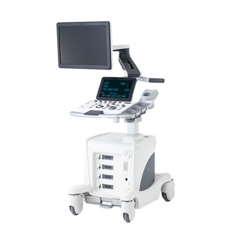
Compact model equipped with a monitor arm for easy operation by both beginners and expert.
This entry model of the ARIETTA series invites easy operation from beginners through experts. Its outstandingly easy-to-view 21.5 inch monitor and intuitive, user-friendly operability will bring you to the “Next Stage”.

Watch the video to know more!
Carefree Workflow
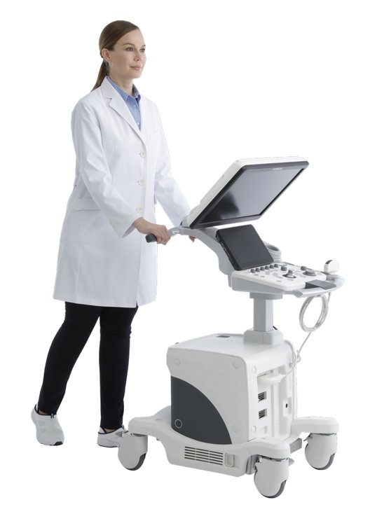
Flexible Monitor Arm
The monitor arm can be moved freely to give you greater flexibility when performing multiple styles of exams.

Easy-to-use Touch Screen Panel
The touch screen panel is mounted at a comfortable and convenient angle. The screen’s layout is customizable by clinical application, allowing for intuitive operation.
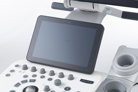
Adjustable Panel Height/ Rotation
It is possible to adjust the height and rotation of the operating console to best suit the operator and examination to be performed.
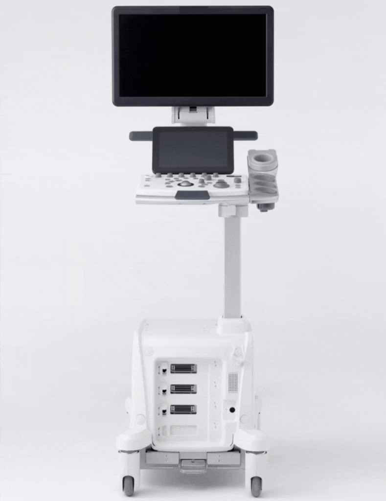
21.5 inch Widescreen Monitor
The high contrast LCD monitor with a wide field of view displays images with high sensitivity and resolution, reducing patient-dependent image variability.

Simplified Operating Console
Minimalized layout includes only the necessary controls. Reduces time searching for the right button, offering a more pleasant operating experience.

Battery*
Battery operation enables the system to be moved to a new location without powering it down, so that operation can be resumed immediately.
*1 The standard components and optional items differ depending on the country.
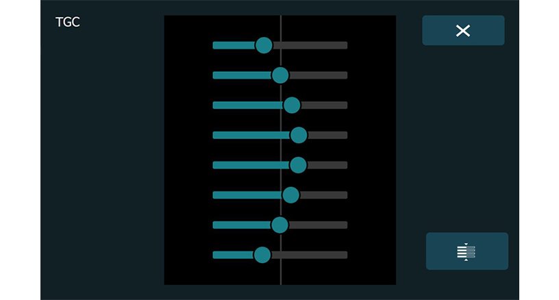
Virtual TGC
Intuitive and smooth operations are possible.
User-friendly Interface
Select an examination area from the illustration to start operation.
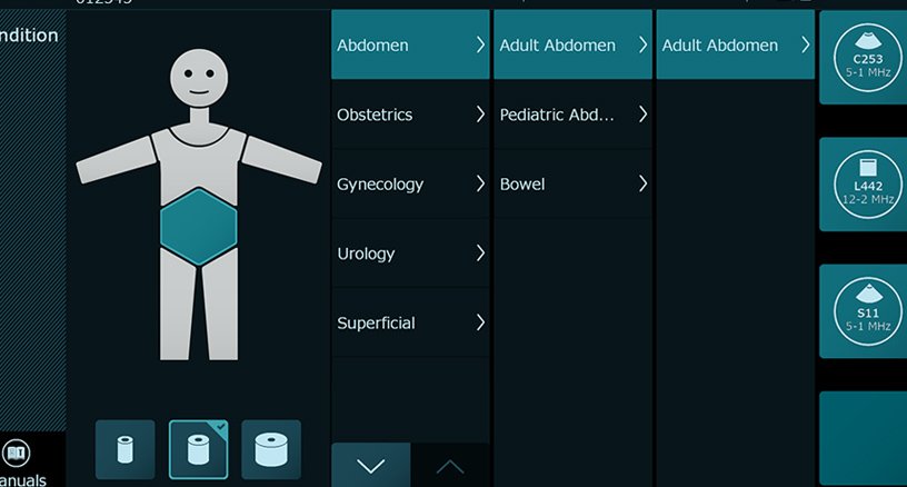
Scan Using a Previous Setting
Choose a patient’s previously-acquired image and the system will adjust to the same exam settings; invaluable for follow-up comparisons.

Clear Imaging
Pure Symphonic Architecture
Technology to support diagnostic imaging
The technologies committed to create the high quality “sound”, which have been fostered by the ARIETTA brand, are inherited.

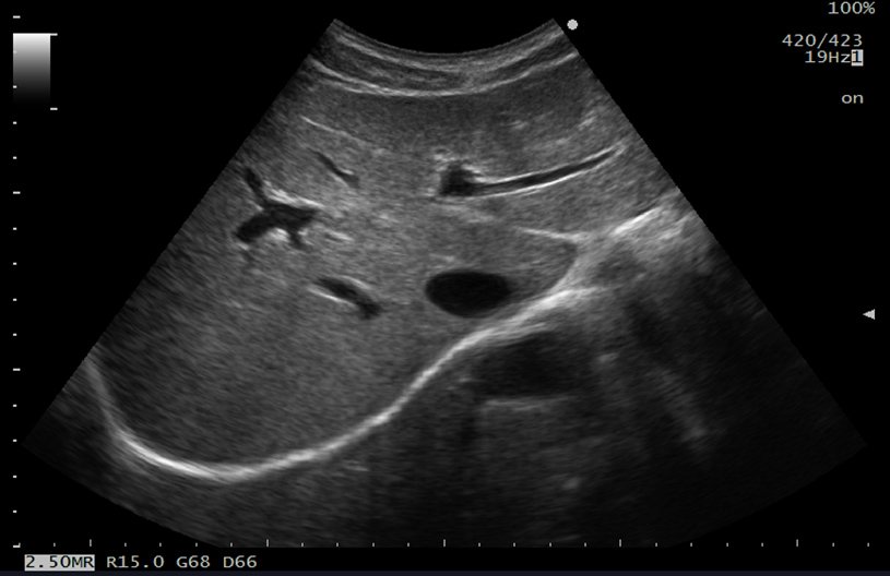
Silky Image Processing (SIP)
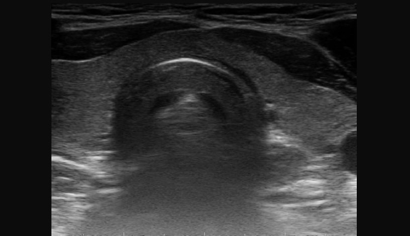
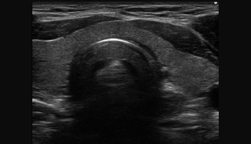
This adaptive image filter has further evolved to reduce speckles and boost edge enhancement, generating clearer images for easier interpretation.
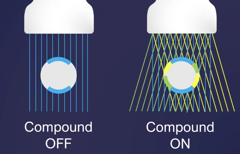
Compound Imaging
Enhances visualization of tissue boundaries and reduces artifacts. Ultrasound beams are transmitted in multiple directions to scan the object from different angles. These scans are superimposed in real time, improving contrast resolution and reducing speckles, thus allowing clearer observation of lesions.
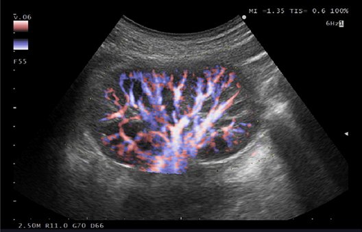
eFLOW
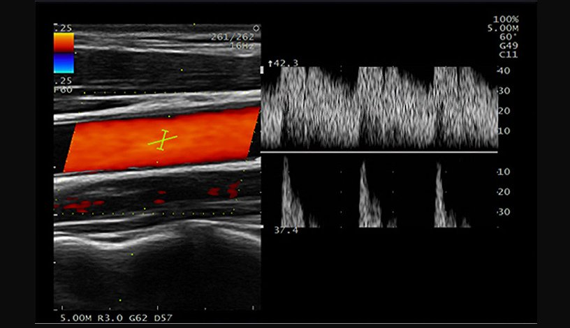
eFLOW is a flow mapping technology that enables accurate and detailed depiction of blood flow dynamics.
Its exceptional spatial resolution allows accurate delineation of both fine and larger blood vessels.
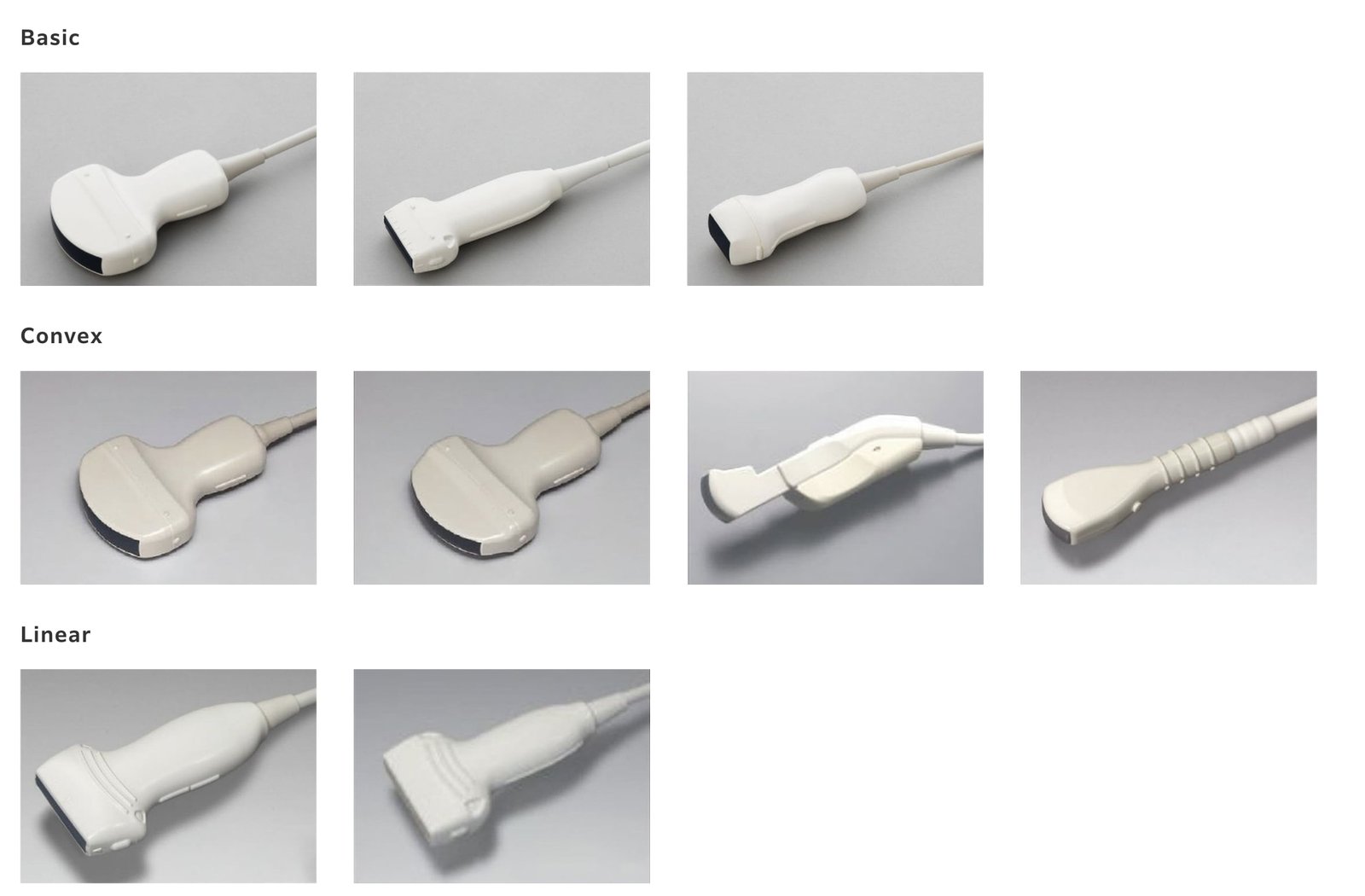
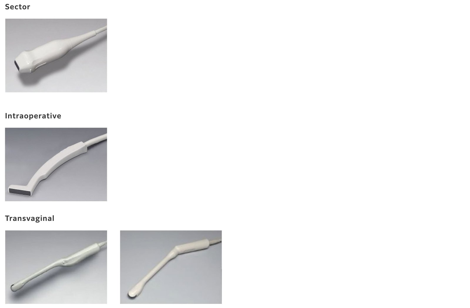
Transducer line-up
Supports a comprehensive range of transducers that support a diverse clinical application. Compatible with a line of newly developed transducers, as well as a selection of transducers from our high-performing systems and other systems.

Clean Application
Trapezoidal Scanning
Offers a wider field of view when scanning with linear transducers, enhancing the visualization of vessels, organs, and the tissues surrounding them.

Auto IMT
Automatically measures the max and mean values of Intima-Media Thickness (IMT) following the placement of an ROI (Region of interest) on the long axis view of the carotid artery.
Because it is calculated from all the points in the specified range, improvement in accuracy can be expected.

Dynamic Slow-motion Display (D.S.D)
D.S.D. displays a real-time image and its slow-motion counterpart side by side on one screen. Rapid valve movement can be observed in detail.
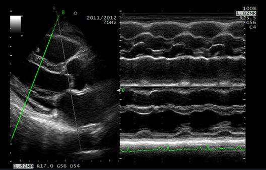
Free Angular M-mode (FAM)
The M-mode can be displayed using any cursor orientation.
Wall and valve movement can be compared from multiple angles in the same heartbeat.
Doppler Auto Trace
Follows the Doppler waveform in real time and displays the measured value. Since freezing and peak velocity and blood vessel resistance value (PI, RI) are displayed at the same time, it is possible take measurements faster.


